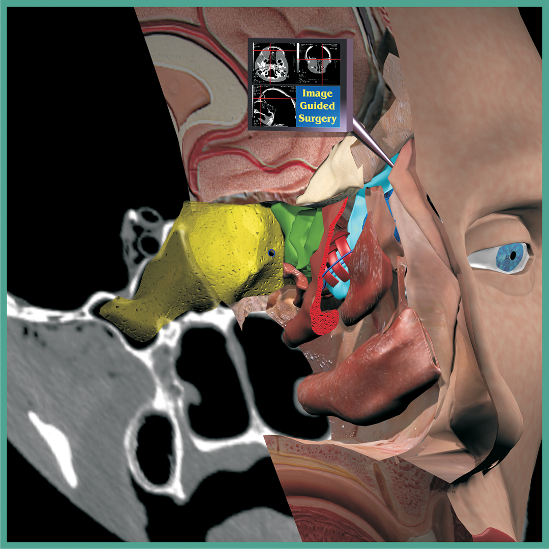
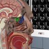
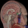
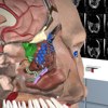
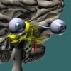
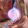
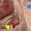
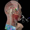
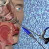
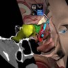
This series of pictures only shows simple anatomy. In my advanced lectures for sinus surgeons, more complex topics can be covered. Here you can see an image-guided surgery system, similar to the kind used during real endoscopic operations. Wherever the pointer touches, the matching CAT scan slices are shown on screen. The CAT scan data can also be dropped right into the 3D scene, as shown here. The scan at the level of the sphenoid sinus is visible. The right sphenoid is partly see-through. A small anatomic landmark called the hiatus semilunaris superior is also pointed out.

Introduction 1

The Lab

Sinuses Side
Sinuses Front
Mid-Sagittal
Septum
Deviated Septum
Deviated Septum

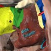
Inferior Turbinate
Inferior Turbinate
Middle Turbinate
Concha Bullosa
Middle Turbinate
Middle Turbinate
Airflow
Airflow
Frontal
Frontal
Drainage
Drainage
Drainage
Drainage
Sphenoid
Sphenoid

Maxillary
Maxillary
Maxillary

Mucous Flow
Ostio-Meatal
Ostio-Meatal
Polyps
Polyps
Windows
Windows
Endoscopic
Endoscopic
Turbinate Reduction

Turbinate Reduction
Cool Picture